

How are Ultrasound Tests Performed for Cancer Detection?
Key Takeaways
Ultrasound tests for cancer detection use transducers to transmit sound waves that create images distinguishing healthy from abnormal tissue, with some tumors requiring invasive probes and Doppler flow machines to assess blood vessel involvement. Procedures typically last 20-40 minutes and can be followed by ultrasound-guided biopsies to examine suspicious tissue.
- Acoustic gel and transducer pressed against skin transmits sound waves internally
- Echoes create real-time images showing tissue differences between healthy and abnormal masses
- Invasive ultrasound uses probes inserted into body to detect prostate or ovary tumors
- Abnormal masses appear light-colored vs. typical black for benign fluid-filled cysts
- Doppler flow machines assess blood vessel involvement; biopsy can pinpoint tissue for examination
If you have compassion and a desire to help others, then the medical field may be the ideal career for you. There are numerous specialties to choose from, but if your passion is to work with patients hands-on, then consider becoming an ultrasound technician.
Commonly known for maternal health, ultrasound tests are also performed for cancer detection. This is where not only your skills are needed, but also your natural concern for others. Because this test uses no radiation and is typically cheaper for patients, it is often one of the first imaging tests used when on the road to a diagnosis.
How are Ultrasound Tests Performed for Cancer Detection?
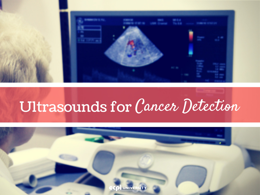
Ultrasounds can be used to detect cancer in various parts of the body, such as the breasts, liver, kidneys, and pancreas. Also called sonography, this imaging test uses high-frequency sound waves to create detailed pictures of the patient’s internal organs and tissues.
To do so, the ultrasound technician first applies acoustic gel to the skin. Then a sonar-like transducer is pressed against and moved along the skin surface to transmit sound waves internally. As the sound waves hit the internal organs and tissues, the echoes bounce back to the device. These echoes are then analyzed by the computer and displayed on the screen in either a conventional flat format or as a 3-D or 4-D image.
Although many abnormalities can be located by external use of the transducer, others require an invasive ultrasound. To detect and locate tumors in the prostate or ovaries, the technician uses a sonography probe that is inserted into the body.
The echoes produced by the transducer then create a real-life picture on the screen, which differs in appearance between healthy and unhealthy tissue. For example, when the sound waves hit an abnormal mass they will appear light-colored, instead of the black sound waves typically produced by fluid-filled, and often benign, cysts.
Ultrasound tests performed for cancer detection typically take 20 to 40 minutes, depending on the location and depth of the organ being scanned. Once complete, the images are interpreted and the results are sent back to the physician.
How are Ultrasounds used after an Abnormality is Detected?
If a tumor or other mass is found, an ultrasound-guided biopsy can help pinpoint the exact location of the tissue needing to be examined. In case of a cancer diagnosis, the ultrasound becomes an important tool for monitoring the tumor for changes in size and to detect if the cancer has spread to other organs.
Additionally, to see if the cancer has spread into the blood vessels, the technician may use a Doppler flow machine which can show any abnormality in the speed or direction of blood flow.
What do you need to know to Effectively Deal with People Facing a Cancer Diagnosis?
Compassion is a necessity when doing ultrasound tests performed for cancer detection. As the technician, you are the first person that patients will look to for information. They will likely be nervous, afraid and perhaps quite emotional, and it is up to you to try to calm them.
From the moment they enter the room, use your voice and body language to create a warm and comforting atmosphere. Carefully explain to them how the process works and give them an opportunity to ask questions. If possible, make friendly conversation to help them focus their mind on something other than the test.
The patient’s calmness is important both for their own peace of mind, as well as the imaging test itself. For optimal accuracy, the patient needs to be as still as possible during the scan and capable of moving and breathing as instructed.
Education can Prepare you for Being a Medical Sonographer
The job of an ultrasound technician requires exceptional technical skills, as well as an array of personal attributes. Fortunately, all the tools you need in order to become a successful and trusted technician can be achieved through a Diagnostic Medical Sonography degree program.
The standard curriculum covers everything from instrumentation to psychology. You will learn the skills of communication, problem solving and critical thinking. You will also have the opportunity to obtain on-the-field clinical experience, which will help you to perform routine sonographic procedures, while simultaneously building your confidence to do so.
Are you interested in a rewarding career as an ultrasound technician? If so, contact ECPI University today for more information about earning your Associate of Applied Science in Diagnostic Medical Sonography. Admissions advisors are waiting to speak to you today about your future in the health science field.
It could be the Best Decision You Ever Make!

DISCLAIMER – ECPI University makes no claim, warranty, or guarantee as to actual employability or earning potential to current, past or future students or graduates of any educational program we offer. The ECPI University website is published for informational purposes only. Every effort is made to ensure the accuracy of information contained on the ECPI.edu domain; however, no warranty of accuracy is made. No contractual rights, either expressed or implied, are created by its content.
Gainful Employment Information – Sonography - Associate’s
For more information about ECPI University or any of our programs click here: http://www.ecpi.edu/ or http://ow.ly/Ca1ya.
