

The Brilliant Technology Behind the Invention of the Ultrasound
Key Takeaways
Ultrasound technology evolved from scientific discoveries about echolocation and piezoelectricity into portable medical imaging devices. Pierre Curie's 1877 discovery of piezoelectricity enabled transducers vibrating at ultrasonic frequencies, while Paul Langevin developed sonography in 1915 initially to locate submerged objects after the Titanic sinking.
- Lazzaro Spallanzani proved bats navigate via echolocation in the 1790s, though the discovery was rejected for nearly 150 years
- Piezoelectric effect allows transducers to vibrate at ultrasonic frequencies for medical imaging
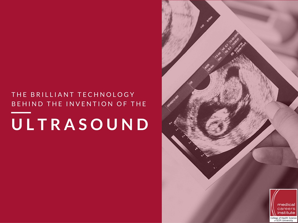
- Sound waves are converted into visible light images for real-time soft tissue assessment
- Modern ultrasound machines range from room-sized equipment to handheld portable devices
- Portable ultrasound enables diverse applications from sports medicine to zoo animal health monitoring
The emerald is rarer than a diamond. It is an exquisite green, more precious by far than many other gemstones. It is a beryl, in Latin beryllus, from which we get the word “brilliant,” meaning shiny and radiant. Ultrasound itself is, at its literal core, in no way “brilliant,” for the simple reason that sound waves are not light waves. We need to combine the breathtaking technology of ultrasound with the literal brilliance of CRT screens to provide an unsurpassed tool for medical diagnosis. The thinking that created the technology behind ultrasound is truly brilliant, though. Ultrasound is marvelous for its ability to peer inside patients with nary a needle or knife to be seen.
The Invention of the Ultrasound
Like most inventions, ultrasound has many fathers. The source of inspiration may have been bats, or icebergs, or soccer. It depends on the strand you grasp onto and unravel from the entire skein of development.
Lazzaro Spallanzani tried in the 1790s to find out how blindfolded bats could still navigate (just imagine for a moment this Italian priest blindfolding bats). He brilliantly realized their ears were more efficient than their eyes, a stance firmly rejected by leading scientists until 1938, when he was shown to be right all along. Spallanzani could not be reached for comment about being vindicated, having died in 1799.
In 1877, Pierre Curie discovered piezoelectricity, which allowed a transducer to vibrate at ultrasonic frequencies. This led to sonographic imaging by Paul Langevin in 1915. He was attempting to hunt down submerged objects in the wake of the Titanic’s 1912 sinking.
Medical Ultrasound
Doctors always have patients’ best interests at heart, so they naturally seized upon ultrasound as a breakthrough in treating...well, just about everything. European soccer teams received physical therapy with ultrasound in the 1920s and 1930s. Gastric ulcers and eczema were subjected to ultrasonic waves. In 1942, Karl Dussik used ultrasound in attempting to find brain tumors. John Wild used amplitude-mode (A-mode) and brightness mode (B-mode) instruments to hunt for intestinal and breast malignancies in 1955.
Ian Donald developed a two-dimensional scanner and an automatic scanner in 1960, leading to early pregnancy detection (six to seven weeks) in 1963. In the early 1970s ultrasound was used successfully on musculoskeletal features, building on Dussik’s 1958 work.
How Does an Ultrasound Work?
From large machines that overwhelmed entire rooms, ultrasound machines today can fit in the palm of your hand. Portable devices proliferate, allowing trainers to immediately assess sports injuries, zookeepers to check animals’ health on the hoof, and skittish patients to relax in a chair while a sonographic technician checks them in complete comfort.
One major manufacturer, Philips, provides machines for both diagnosis and intervention:
- Breast Imaging
- Cardiology
- Hepatology
- Internal Medicine
- Vascular
- Musculoskeletal
- Obstetrics and gynecology
- Radiology
Remembering that sonography depends on sound waves, these technologically advanced machines can provide extremely accurate images in which the sound waves are interpreted and displayed as visible light, providing medical professionals immediate feedback on soft tissue conditions inside a patient. Doctors can see more clearly, with minimal patient discomfort, than ever before.
Coupled with powerful computers, ultrasound machines can generate applications such as Philips’ HeartModel, an Anatomically Intelligent Ultrasound (AIUS) bringing live 3D quantification and automated 2D views to echocardiography. Cardiologists, working with experienced sonographers, can immediately learn heart ventricle volumes, heart health, and “see” 3D images to make quick decisions about patients.
Earn a Degree in Diagnostic Medical Sonography
You may not be able to hear the ultrasounds these amazing machines produce, but you should listen to this bit of advice. You can get an excellent education that values the long tradition of ultrasound while teaching leading-edge techniques on modern equipment. Attend ECPI University to earn your Associate of Applied Science in Diagnostic Medical Sonography in around 18 months. Graduate, take examinations in ultrasound specialities, and then employers will be all ears, ready to hear from you. Contact ECPI University today to hear how your future as a diagnostic medical sonographer could be brilliant—it could be the Best Decision You Ever Make!
Ultrasound technology at ecpi is what I plan to do
— Ana (@anaa_preciado) October 2, 2014

DISCLAIMER – ECPI University makes no claim, warranty, or guarantee as to actual employability or earning potential to current, past or future students or graduates of any educational program we offer. The ECPI University website is published for informational purposes only. Every effort is made to ensure the accuracy of information contained on the ECPI.edu domain; however, no warranty of accuracy is made. No contractual rights, either expressed or implied, are created by its content.
For more information about ECPI University or any of our programs click here: http://www.ecpi.edu/ or http://ow.ly/Ca1ya.
